File:Fig1 Clay CancerInformatics2017 16.png
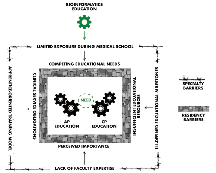
Fig1_Clay_CancerInformatics2017_16.png (718 × 576 pixels, file size: 147 KB, MIME type: image/png)
Summary
| Description |
Figure 1. Unique barriers to implement bioinformatics education into pathology residency training programs. Effective bioinformatics education could integrate into both anatomic pathology (AP) and clinical pathology (CP) and function to facilitate learning in both disciplines. A few of the most relevant barriers are illustrated here. The outer fence represents some of the challenges that are unique to the specialty of pathology. The arrows indicate how these challenges manifest at the residency training level and act as barriers (denoted by the inner wall). |
|---|---|
| Source |
Clay, M.R.; Fisher, K.E. (2017). "Bioinformatics education in pathology training: Current scope and future direction". Cancer Informatics 16: 1–6. doi:10.1177/1176935117703389. PMC PMC5392012. PMID 28469393. http://www.pubmedcentral.nih.gov/articlerender.fcgi?tool=pmcentrez&artid=PMC5392012. |
| Date |
2017 |
| Author |
Clay, M.R.; Fisher, K.E. |
| Permission (Reusing this file) |
Creative Commons Attribution-NonCommercial 4.0 International |
| Other versions |
Licensing
|
|
This work is licensed under the Creative Commons Attribution-NonCommercial 4.0 International License. |
File history
Click on a date/time to view the file as it appeared at that time.
| Date/Time | Thumbnail | Dimensions | User | Comment | |
|---|---|---|---|---|---|
| current | 17:25, 9 August 2017 | | 718 × 576 (147 KB) | Shawndouglas (talk | contribs) |
You cannot overwrite this file.
File usage
The following 2 pages use this file:


