File:Fig2 Wang BMCBioinfo2019 20.png
Original file (1,946 × 1,112 pixels, file size: 266 KB, MIME type: image/png)
Summary
| Description |
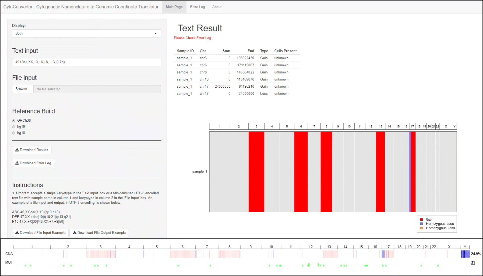
Fig. 2 CytoConverter output from a cancer cell line. The top panel shows a screenshot of CytoConverter as applied to the karyotype of the cell line AML-193. The output produced by our web tool is under “Text Result,” wherein the sample is referred to as “sample_1” by default. The bottom panel shows the results from microarray-based copy number analysis of AML-193, as performed in Barretina et al. and visualized using cBioPortal. In cBioPortal output, red indicates gains, blue indicates losses, and a darker color indicates a larger deviation from normal (i.e., a “deeper” deletion or higher-level amplification). Note that cBioPortal reports Y as having been lost, though the cell line is derived from a male patient with only X chromosomes constitutionally and therefore the Y chromosome was not present in the sample. |
|---|---|
| Source |
Wang, J.; LaFramboise, T. (2019). "CytoConverter: A web-based tool to convert karyotypes to genomic coordinates". BMC Bioinformatics 20: 467. doi:10.1186/s12859-019-3062-4. |
| Date |
2019 |
| Author |
Wang, J.; LaFramboise, T. |
| Permission (Reusing this file) |
|
| Other versions |
Licensing
|
|
This work is licensed under the Creative Commons Attribution 4.0 License. |
File history
Click on a date/time to view the file as it appeared at that time.
| Date/Time | Thumbnail | Dimensions | User | Comment | |
|---|---|---|---|---|---|
| current | 22:48, 16 December 2019 | | 1,946 × 1,112 (266 KB) | Shawndouglas (talk | contribs) |
You cannot overwrite this file.
File usage
The following page uses this file:


